Research

Molecular factors controlling human brain development
Our lab has a long-standing interest in studying the pathways controlling regionalisation and specification of the human developing brain. This work involves analysis of fetal brain anatomy, and identification of key genes and noncoding RNAs controlling the compartmentalisation of the brain. Through the use of human embryonic stem cells (hESCs), we can mimic brain development towards different regions of the human brain, and thereby investigate the effect of novel genes on neural differentiation.
We also work on more sophisticated 3D culturing methods to model human brain development on an anatomical level with hESCs. In this project, involving engineers from LTH, we apply advanced microfluidic techniques to culture hESCs under the influence of chemical gradients to mimic the environment around the developing brain in the fetus, thereby generating neural tissue with anatomical characteristics resembling the developing human brain.

Developing human dopamine neurons for clinical translation
As we have a strong focus on developing cells for cell replacement therapy in Parkinson’s disease, we are currently adapting our protocols for GMP compliance through the EU-funded network NeuroStemcellRepair. In line with this aim, we also perform extensive preclinical validation of the human dopamine neurons derived from our protocol in rat models of Parkinson’s disease. This includes long-term survival of transplanted cells in vivo to assess cell fate, maturation and integration, as well as functional behavioural assessment for graft function in vivo.
As a member of the European clinical trial TRANSEURO we also perform preclinical validation of human fetal dopamine neurons for clinical use. This also allows us the unique opportunity to directly compare our hESC-derived dopamine neurons with those sourced from human fetal tissue both in vitro and in vivo.

New methodologies for in vivo assessment of human neurons generated from stem cells or via reprogramming
We monitor our cells after transplantation using standard in vivo assessments as listed above. However, with a growing number of novel cell sources (stem cells and reprogrammed cells) it becomes important to develop new methodologies for a more thorough assessment of human neurons generated using these approaches. Thus we have recently established a number of new technologies to reveal how transplants function and integrate with the host brain.
We utilise monosynaptic tracing technology, using pseudotyped rabies vectors, in order to visualise the synaptic contacts formed between host neurons and graft human cells. This approach has opened up for a number of possibilities to assess graft integration in vivo that has so far been relatively unaddressed in the field. While expanding our knowledge of graft integration, we also investigate how that occurs at a functional level by utilising optogenetics in combination with highly sensitive and quantitative methods assessing neuronal function, such as electrophysiology and in vivo amperometry.

Direct conversion of human fibroblasts into subtype-specific, functional neurons
We have shown that human fibroblasts can be directly converted into functional and subtype-specific neurons using defined sets of transcription factors. The cells are called induced neurons (iNs) and offer a shortcut over induced pluripotent stem cells (iPSc) for generating patient and disease-specific neurons. We are currently focusing on improving the yield and transplantability of directly reprogrammed dopamine neurons, and also developing reprogramming methodology that are compatible with clinical use.

In vivo neural conversion
Using a transgenic mouse model to specifically direct expression of reprogramming genes to parenchymal astrocytes residing in the striatum, we were recently able to show that endogenous mouse astrocytes can be directly converted into neurons in situ. This study provided important proof-of-principle that direct neural conversion of endogenous glia can take place in the adult rodent brain. Current efforts focus on identifying the optimal starting cell population for neural conversion, identification of gene combinations that yield subtype specificity and analysis of function and integration of the reprogrammed neurons.
This project was funded via an ERC starting grant awarded to M Parmar.
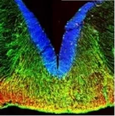
Human midbrain development
We have recently shown that many of the key transcription factors involved in dopamine neuron specification in the rodent brain are expressed in similar spatio-temporal expression domains also during human midbrain development and that it is the radial glia cells located in the floor plate that gives rise to DA neurons also in the human brain. Current projects include a continued mapping of the dopamine domain during human fetal development as well as studies of location of serotonergic neurons and their precursors in the abutting regions of the hindbrain.